Branch Retinal Vein Occlusion (BRVO)

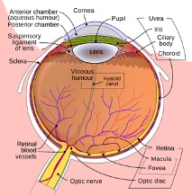
Illustration of ocular circulation.
Just like any other tissue in your body, the retina has a blood supply. The central retinal artery, travels down the optic nerve and supplies blood to the entire retina. At the point the central retinal artery enters the retina, it begins to progressively branch into smaller arteries and then microscopic capillaries, which supply oxygen and nutrients to the retina. This arterial system connects to a parallel set of veins that drain into larger veins leading to the central retinal vein, which exits the retina also through the center of the optic nerve. Some of the most common problems a retina specialist sees are related to abnormalities in the blood vessel system of the retina.
A branch retinal vein occlusion is a blockage of a branch of the central retinal vein. Some branch vein occlusions may be mild, cause little or no vision loss and not require treatment. However, more severe cases may lead to impaired retinal blood supply, retinal hemorrhages and retinal swelling (edema) that can cause loss of vision. Unfortunately, there is no way to reverse a blocked vein. Medications, which must be injected into the eye periodically, may improve vision. The injections may be needed as frequently as once a month, but later on the treatment interval can be extended, and in some cases, treatment may be stopped. Laser is an alternative form of treatment which has been used for many years.
Branch retinal vein occlusions are associated with other systemic medical conditions, such as high blood pressure, high cholesterol, diabetes, heart disease and stroke. Cigarette smoking is also a risk factor. Changes in blood vessels which occur with aging are also a factor. Rarely, branch vein occlusions can be associated with blood clotting disorders. In some cases, branch vein occlusion occurs for no apparent reason. Think of it as a kink in a garden hose.

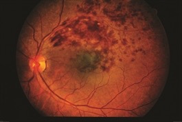
Image of a branch retinal vein occlusion.
Branch vein occlusion usually presents with sudden onset of blurred vision. Eye examination shows retinal hemorrhages and swelling as well as the location of the blockage. Retinal imaging with fluorescein angiograpy and optical coherence tomography (OCT) are helpful. Overall, BRVO carries a generally good prognosis. Over 60% of patients, treated and untreated, maintain vision better than 20/40 after 1 year.