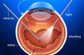
Vitrectomy

Vitrectomy is the most common surgical procedure performed by retina specialists. It is used to treat a variety of retinal conditions including retinal detachment, complications of diabetic retinopathy, macular holes, epiretinal membranes, vitreous hemorrhages, infections, and ocular trauma. A variety of ancillary procedures can be performed along with vitrectomy including laser photocoagulation, scleral buckling, cryosurgery, gas/fluid exchange, and lens removal. The surgery is performed with an operating microscope, using a contact lens and a dilated pupil. Instruments are inserted through tiny openings in the sclera, the white part of the eye. Pressure in the eye is maintained by infusing fluid. Illumination is achieved using a fiberoptic light.
Surgery is typically performed as an outpatient, with local anesthesia. Vital signs are monitored by an anesthesia team. IV sedation can be used to keep patients comfortable and relaxed. A post-op visit is scheduled for the following day where the patch is removed and eyedrops are started. An air or gas bubble is sometimes used to keep the retina in place temporally. There may also be the need to position for a few days after the surgery. There is usually very little pain, and surgical recovery is fairly quick. Most patients can resume normal activity including work, exercise and driving within a week.